
Automated Refraction System
No more clicking lenses back and forth. Precise prescriptions in half the time.
Eye Health Technology
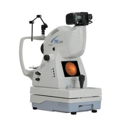
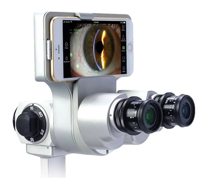
Digital Retinal Photography
The retinal camera records a detailed view of the retina and is a great way to monitor the progression of diabetic retinopathy and ocular tumors.
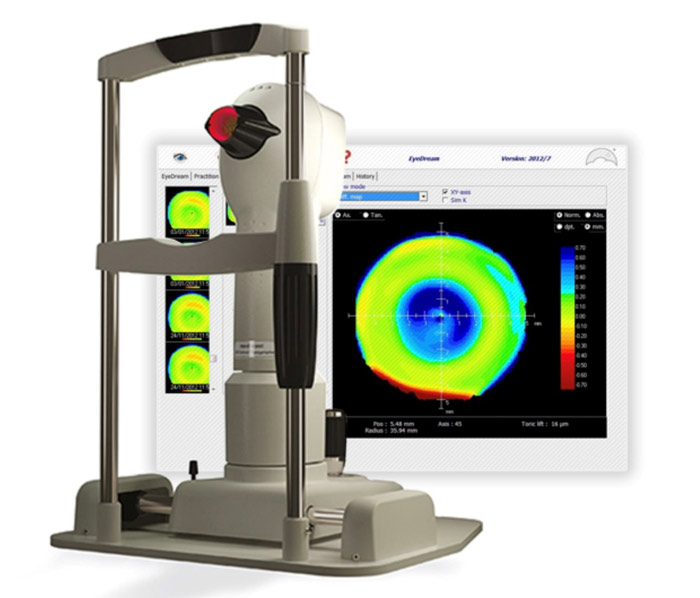
Corneal Topography
The corneal topographer provides a three-dimensional map of the surface of your eye, allowing your doctor to detect corneal conditions such as keratoconus, obtain measurements for refractive surgery, and design specialty contacts for irregular and post-surgical corneas.
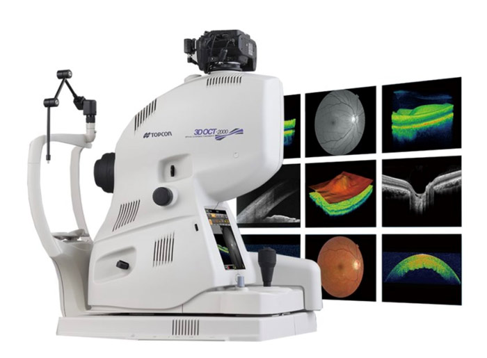
Optical Coherence Tomography (OCT)
The OCT uses light waves to allow your doctor to see the ten layers of the retina which is only 1 mm thick. The device has revolutionized the detection and monitoring of glaucoma and retinal disease.
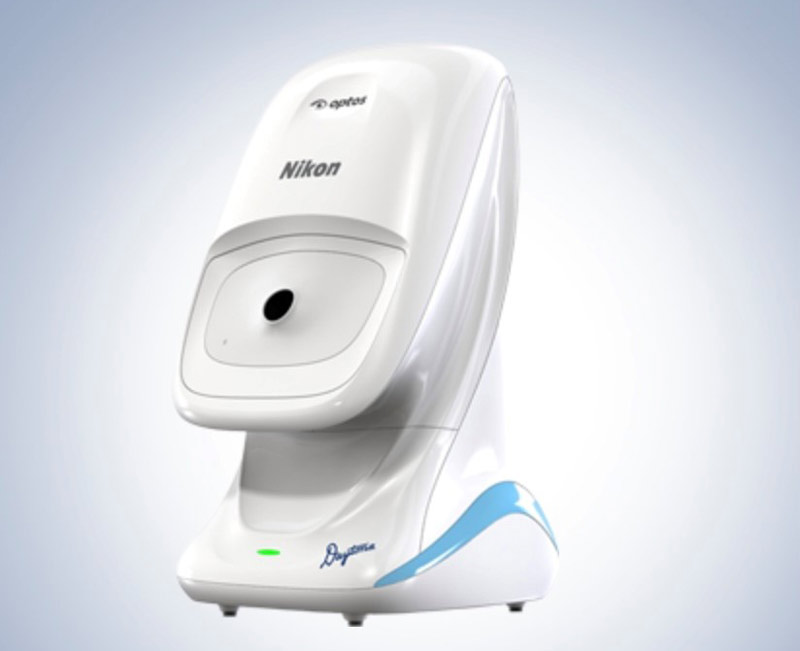
Daytona Optomap Ultra-Widefield Retinal Imaging
The Optos Daytona produces a 200° image of the retina in half a second, allowing visualization of 90% of the retina in an undilated eye.
Visual Field Analyzer
The visual field analyzer is used to detect and monitor glaucoma, neurological disease, and droopy eyelids for cosmetic surgery. The device statistically analyzes the results and compares them to a normative database.
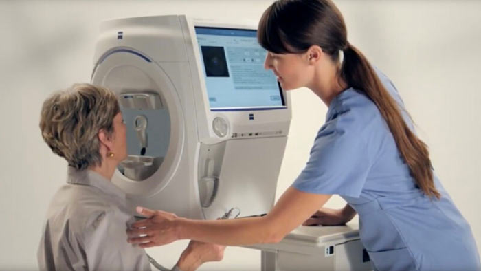
Anterior Segment Imaging
The anterior segment imaging system allows the doctor to monitor any changes on the surface of your eye and is a great educational tool for the patient as the patient can see what the doctor is observing.